Mast Cell Tumors in Dogs: Signs and Treatments
Blog Summary:
Mast cell tumors (MCTs) are among the most common skin cancers in dogs, with behaviors that range from relatively mild to highly aggressive and life-threatening. This blog explores the biology of mast cell tumors, how they develop, and the breeds most often affected. It outlines the clinical signs pet owners may notice such as lumps, changes in size, redness, ulceration, or systemic illness and explains why prompt veterinary evaluation is so important.
The article highlights the advanced diagnostic process offered at Veterinary Specialty Center in Bannockburn, IL, including fine needle aspiration, biopsy and grading, staging tests, and imaging, all of which help determine the tumor’s aggressiveness and spread. Treatment options are discussed in depth, from surgery and radiation therapy to chemotherapy, targeted drugs, and supportive care, with emphasis on tailoring strategies to the individual patient.
Finally, the blog addresses the factors that influence prognosis such as tumor grade, stage, growth rate, and surgical margins and underscores the value of a collaborative, specialty-level approach. Pet parents will come away with a clear understanding of what mast cell tumors are, how they are diagnosed, the treatments available, and what influences long-term outcomes for affected dogs.
Introduction:
A lump on your dog can raise immediate concern—especially when it begins to grow or change in appearance. Among the myriad possibilities, mast cell tumors (MCTs) represent a prevalent and clinically significant cutaneous neoplasm in dogs, characterized by highly variable biological behavior. Their spectrum ranges from indolent, locally contained lesions to aggressive, metastatic disease, underscoring the critical need for precise diagnosis and tailored therapeutic strategies.
At Veterinary Specialty Center in Bannockburn, IL, our collaborative oncology and surgery departments are dedicated to the comprehensive evaluation and management of canine mast cell tumors, employing an evidence-based and individualized approach. This discourse will delineate the typical clinical presentations, advanced diagnostic protocols, and state-of-the-art treatment modalities available through specialized veterinary care for this complex canine neoplasm.
The Biology of Canine Mast Cell Tumors
Mast cell tumors originate from mastocytes, integral components of the immune system responsible for mediating allergic reactions and inflammatory responses. These ubiquitous cells are distributed throughout various bodily tissues. When uncontrolled, they can give rise to cancerous growths, most commonly observed in or immediately beneath the skin. However, mast cell tumors can also manifest in visceral organs such as the spleen, liver, gastrointestinal tract, or bone marrow.
The inherent unpredictability of canine mast cell tumors is a defining characteristic. While some remain localized and exhibit favorable responses to primary surgical intervention, others demonstrate aggressive local infiltration and a propensity for metastasis to regional lymph nodes, distant organs (e.g., liver, spleen), or bone marrow. This biological variability necessitates early diagnosis and accurate staging to formulate an effective and prognostic treatment plan.
Breed Predispositions for Mast Cell Tumor Development
While mast cell tumors can affect any canine breed, certain genetic predispositions have been identified, leading to a higher incidence in specific breeds. Awareness of these predispositions allows for heightened vigilance by both owners and veterinary practitioners for early detection of suspicious lesions.
Breeds Commonly Affected
- Boxers
- Boston Terriers
- Bulldogs (English and French)
- Pugs
- Labrador Retrievers
- Golden Retrievers
- Shar-Peis
It is noteworthy that breeds like Boxers and Pugs frequently present with lower-grade, less aggressive forms of MCTs. Conversely, Shar-Peis are often predisposed to high-grade variants that typically mandate more intensive and multimodal therapeutic interventions.
Clinical Presentation of Canine Mast Cell Tumors
The clinical presentation of mast cell tumors is remarkably diverse, often posing a diagnostic challenge without definitive cytological or histopathological evaluation. Although the majority are cutaneous, deeper subcutaneous or visceral involvement can also occur.
Key Clinical Signs to Observe
- Palpable Mass: A raised, firm, or sometimes pliable lump or nodule on or beneath the skin.
- Fluctuating Size: Intermittent swelling or shrinking of the mass, often termed “Darier’s sign,” can occur due to mast cell degranulation and subsequent histamine release.
- Local Inflammation: Erythema (redness), pruritus (itching), or irritation of the skin adjacent to the growth.
- Ulceration/Bleeding: The surface of the tumor may become ulcerated or bleed, particularly with trauma or rapid growth.
- Systemic Signs (with Visceral Involvement or Degranulation): Vomiting, diarrhea, anorexia, or lethargy may indicate visceral involvement (e.g., gastrointestinal tract) or systemic effects of massive histamine release.
The release of histamine and other inflammatory mediators by mast cell tumors can induce localized edema and, in some cases, severe gastrointestinal ulceration. Any newly identified or changing mass warrants prompt professional veterinary evaluation.
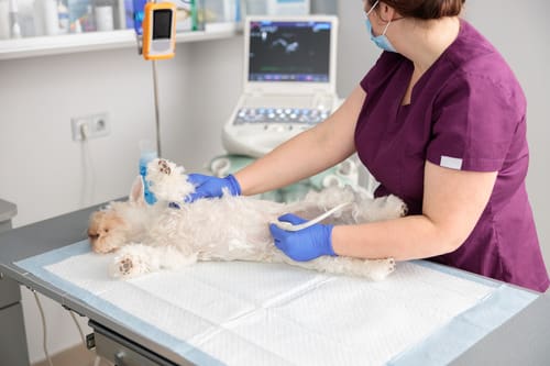
Advanced Diagnostic Approach at a Specialty Center
At Veterinary Specialty Center, our diagnostic protocol for suspected mast cell tumors is meticulously designed to provide a comprehensive understanding of the tumor’s biological behavior and metastatic potential. A collaborative effort among our surgical, medical oncology, radiation oncology, and diagnostic imaging teams ensures a thorough and individualized diagnostic workup.
Fine Needle Aspiration (FNA) and Cytology
Fine needle aspiration is a minimally invasive initial diagnostic tool used to collect cellular material from the mass. Cytological examination of these samples can frequently identify the characteristic granulated mast cells, providing a presumptive diagnosis and guiding subsequent diagnostic and therapeutic planning.
Biopsy and Histopathology
A surgical biopsy is essential for definitive diagnosis and, critically, for accurate tumor grading. Histopathological assessment of the excised tissue allows for classification of the, which is paramount in predicting the likelihood of local recurrence and distant metastasis. Low-grade tumors generally exhibit less aggressive behavior, while high-grade tumors typically necessitate more aggressive, often multimodal, therapeutic strategies.
Staging Tests
To precisely determine the extent of disease and identify any metastatic spread, a comprehensive staging workup may include:
- Regional Lymph Node Evaluation: Cytological or histopathological examination of draining lymph nodes is critical to detect regional metastasis.
- Abdominal Ultrasonography: To assess for visceral involvement (e.g., liver, spleen, gastrointestinal tract) and evaluate abdominal lymph nodes.
- Thoracic Radiographs (X-rays): To screen for pulmonary metastasis, although lung metastasis from MCTs is less common than from other tumor types.
- Complete Blood Count (CBC) and Serum Biochemistry Profile: To assess overall health, identify any systemic effects, and evaluate organ function.
- Urinalysis: To assess renal function and rule out urinary tract issues.
These diagnostic insights collectively inform the precise extent of the disease, enabling the formulation of the most appropriate and prognostically relevant treatment plan.
Comprehensive Management of Canine Mast Cell Tumors
The treatment of canine mast cell tumors is frequently multimodal, integrating surgery, radiation therapy, chemotherapy, and targeted pharmacotherapy. The optimal therapeutic strategy is tailored to the tumor’s grade, anatomical location, and confirmed stage of disease.
Surgery
Surgical excision remains the cornerstone of treatment for localized mast cell tumors. Our board-certified surgeons strive for wide surgical margins to maximize the likelihood of complete tumor removal and minimize the risk of local recurrence. In cases where tumors are situated in anatomically challenging locations (e.g., muzzle, distal limbs), advanced reconstructive surgical techniques may be employed to achieve adequate margins while preserving function and cosmesis.
Radiation Therapy
For tumors that have been incompletely excised or those located in areas where achieving wide surgical margins is not feasible, radiation therapy offers a highly effective option for achieving local disease control. Our radiation oncology specialists develop meticulously customized protocols based on the tumor’s characteristics, the patient’s overall health, and staging information.
Chemotherapy
High-grade mast cell tumors or those with confirmed regional or distant metastasis often benefit from systemic chemotherapy. Administered under the expert guidance of a residency-trained or board-certified veterinary oncologist, chemotherapy can significantly slow disease progression, mitigate metastatic spread, and enhance the patient’s quality of life. Notably, most canine patients tolerate these chemotherapeutic regimens remarkably well, typically experiencing fewer and less severe side effects compared to human oncology patients.
Targeted Therapies
Certain mast cell tumors may respond favorably to targeted therapies, such as tyrosine kinase inhibitors (e.g., Toceranib phosphate – Palladia®). These innovative drugs specifically interfere with molecular pathways crucial for tumor growth and proliferation. Additionally, supportive medications, including antihistamines (e.g., diphenhydramine, famotidine) and gastroprotectants (e.g., omeprazole, sucralfate), are frequently prescribed to manage clinical signs associated with mast cell degranulation.
Factors Influencing Prognosis
Every case of canine mast cell tumor is unique, and the ultimate prognosis is influenced by a confluence of factors. With timely and appropriate therapeutic intervention, many dogs can achieve prolonged periods of remission and excellent quality of life for months to years following diagnosis.
Key Prognostic Indicators
- Histologic Grade: The most significant prognostic factor, distinguishing between low-grade (more favorable) and high-grade (less favorable) tumors.
- Clinical Stage: The extent of disease spread (e.g., localized, regional lymph node involvement, distant metastasis).
- Tumor Location: Tumors on mucocutaneous junctions, nail beds, or internal organs (e.g., gastrointestinal tract) often carry a less favorable prognosis.
- Growth Rate: Rapidly growing or changing tumors are often more aggressive.
- Surgical Margins: Complete surgical excision with clean margins is associated with a better prognosis for local control.
- Presence of Systemic Signs: Signs related to histamine release or visceral involvement may indicate more aggressive disease.
- Proliferative Index (e.g., Ki-67): Molecular markers providing additional prognostic information.
At Veterinary Specialty Center, we synthesize this critical information to provide clear prognostic expectations and meticulously tailored care recommendations for each individual patient.
Our Collaborative Approach to Canine Mast Cell Tumor Management
Effective management of canine mast cell tumors demands an experienced and highly collaborative team approach. At Veterinary Specialty Center, our specialists operate as a unified entity, meticulously coordinating diagnostics, crafting personalized treatment regimens, and offering unwavering support to both our patients and their families throughout the entire journey. Whether your cherished companion has recently received a diagnosis of a mast cell tumor, or your primary veterinarian has recommended a specialized consultation, our oncology and surgery departments are exceptionally well-equipped to assist.
We are proud to offer advanced diagnostic imaging capabilities, the expertise of board-certified specialists, and an unwavering commitment to compassionate care – delivered with the precision and excellence your pet unequivocally deserves. To schedule a comprehensive consultation, please contact us at (847) 459-7535. Referring veterinarians are encouraged to communicate directly with our specialists to discuss specific cases and facilitate coordinated care.
Recent Posts
About Us
Veterinary Specialty Center is a privately-owned, 24/7 emergency and specialty animal hospital located in Bannockburn, IL. Since 1976, their team of board-certified specialists has delivered advanced, compassionate care, leading the way with innovative treatments and a collaborative approach.