Esophageal Strictures
Esophageal strictures are a band of scar tissue in the esophagus causing a narrowing of the esophagus that makes swallowing, particularly solid food, difficult.


What Causes Esophageal Strictures?
Esophageal strictures occur when there is circumferential damage to the lining of the esophagus, causing it to scar down and narrow the lumen. This can result from reflux, in which acid or bile from the stomach or intestine pools in the esophagus, causing damage. Esophageal strictures also can form after ingestion of a foreign body (such as a bone or rock) that gets stuck in the esophagus, causing damage.
Certain oral medications can increase the risk of esophageal stricture, especially in cats, which is why veterinarians recommend that an oral syringe full of water be given after giving a cat a pill. Rarely, esophageal strictures are congenital and are noted in young dogs and cats when they transition from nursing to solid food.
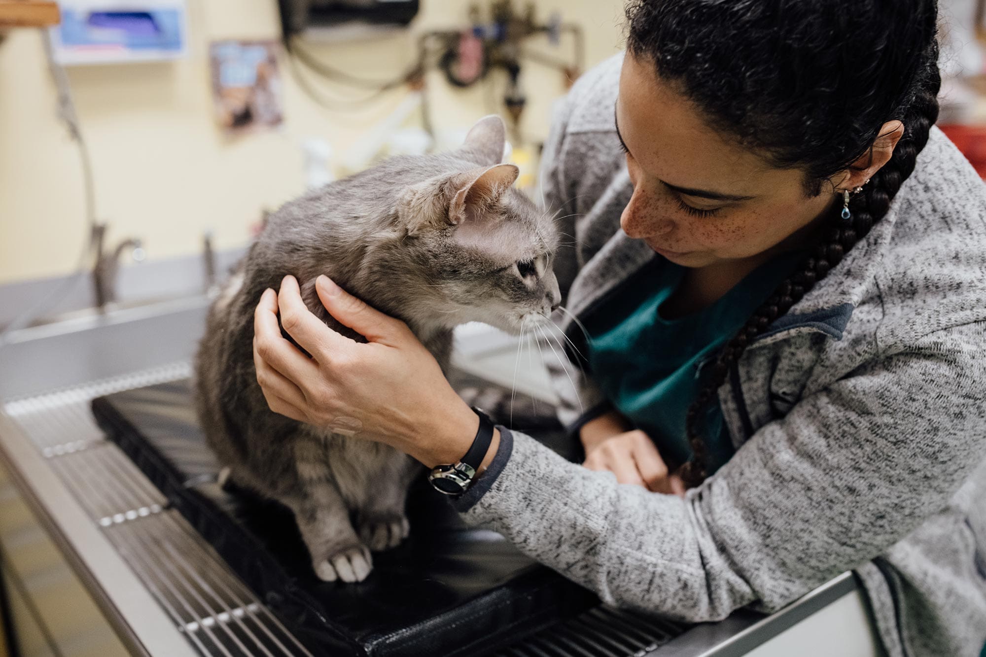
Clinical Signs
Patients with an esophageal stricture generally have a strong desire to eat, but as soon as they attempt to swallow something, they are in immediate distress and appear to be choking until they regurgitate. Soft, liquid food might pass with relative ease, but solid food is difficult or impossible to swallow. In the early phase after esophageal injury, the patient may show distress with swallowing in which they might salivate, foam, and repeatedly make an exaggerated effort to swallow. In time, most patients will regurgitate food immediately after eating. If the problem is not addressed quickly, patients will lose weight and become progressively weaker and more lethargic.


Diagnosis
An esophageal stricture is suspected when a patient has the typical clinical signs of regurgitation, especially when they coincide with a history of recent anesthesia, esophageal foreign body, or oral antibiotic therapy. The diagnosis is generally confirmed with an endoscopic exam of the esophagus. Because patients are often systemically ill and malnourished, additional laboratory tests are done prior to anesthesia for the scope procedure. Radiographs (X-rays) might be done to rule out a tumor in the chest. Because patients with esophageal stricture will regurgitate after eating and sometimes drinking, oral contrast agents such as barium are not recommended as a way to diagnose an esophageal stricture. Doing so puts the patient at risk for aspiration of barium, which can be life-threatening.
Treatment
At this time, the recommended treatment for esophageal strictures is to perform esophageal balloon dilation. In this procedure, a video endoscope is used to visualize the esophageal damage, and an inflatable balloon is passed through the scope and slowly inflated to break down the stricture. This procedure has a general success rate of 70-88% in recent reports with success being defined as accepted gruel or thin foods. It is unlikely these patients will tolerate larger foods such as kibble for the rest of their lives.


A new treatment is under development which incorporates an esophageal feeding tube with an attached balloon. The balloon is inflated at home daily for a period of time and then removed. This allows owners to treat at home and may deliver better success than the traditional endoscopic balloon dilation which is performed every few days in the hospital under anesthesia.
Unfortunately, balloon dilation carries some risks including anesthesia, inflammation of the esophagus (esophagitis), pneumonia due to regurgitation, bleeding, pain, and, in some cases, esophageal tearing. Esophageal tearing is the most concerning and potentially life-threatening complication and this has been documented in 4-9% of cases that undergo this procedure. Some tearing of the esophagus is inevitable to break down the stricture, but if the tear goes too deep, it can rupture the esophagus. To reduce the likelihood that a tear occurs, the stricture ballooning is done very slowly, with progressively larger balloons being passed one after the other.


If a significant esophageal tear is found, emergency surgery may be needed, possibly requiring chest opening. Esophageal strictures often need multiple procedures to restore function, with most patients requiring four to six endoscopic balloon procedures spaced a few days to a week apart. During this time, a feeding tube may be placed for nutritional support. Steroid injections are typically given during the first procedure to prevent scar reformation. Pain medications, antacids, and sometimes anti-inflammatory drugs are prescribed, and a follow-up appointment should be scheduled before leaving.


Prognosis
Esophageal strictures are profoundly difficult to treat and the balloon dilation procedures carry some risk. Patients in which the procedure results in dilation of the esophagus will likely not have a normal life, since they will need to eat a modified diet and cannot tolerate normal dog treats in the future. However, they may still have a good quality of life. Unfortunately, it often takes several procedures to reach that point.
Long Term Follow-Up
Patients with esophageal stricture are treated and followed by the internal medicine specialists at Veterinary Specialty Center. Patients often need multiple procedures as well as supportive therapy such as temporary feeding tubes. Once the patient is able to eat adequately and comfortably orally, feeding tubes are removed and the patients no longer need specialty care.