The larynx, located at the opening of the trachea (windpipe), plays a crucial role in breathing and swallowing. It opens wide to allow air in and closes to prevent food or saliva from entering the trachea and lungs.
Laryngeal paralysis (LP) is a progressive condition most commonly seen in Labrador retrievers and other large breeds, though it can also affect cats. It occurs when the nerves controlling the larynx's opening muscles malfunction. Possible underlying causes include:
-
Hypothyroidism
-
Cervical trauma
-
Thyroid tumors
-
Previous cervical surgeries
-
Neurological diseases
-
Idiopathic
-
Genetics
Breeds such as the Bouvier des Flandres, Siberian husky, dalmatian, rottweiler, American Staffordshire terrier, and black Russian terrier may have hereditary forms of this condition.
Laryngeal paralysis is also often part of a generalized nerve and muscle weakening syndrome called geriatric onset laryngeal paralysis and polyneuropathy (GOLPP).
When laryngeal paralysis occurs, the affected pet struggles to breathe, which can lead to more swelling in the larynx and increased distress, potentially resulting in collapse or cyanosis (turning blue).
Clinical Signs of Laryngeal Paralysis


-
Noisy breathing
-
Excessive panting
-
Exercise intolerance, expecially in warm weather
-
Changes in bark (or meow)
-
Coughing, gagging, or regurgitation while eating or drinking
-
Difficulty breathing
-
Blue tint to tongue or gums
-
Collapse
Diagnosis of Laryngeal Paralysis
Diagnosing laryngeal paralysis requires an examination under sedation to assess laryngeal function. Additional tests, such as chest and neck X-rays, bloodwork (including thyroid levels), and a thorough neck examination, may be necessary to rule out other health issues.
Surgical Treatment of
Laryngeal Paralysis
Surgical Treatment of Laryngeal Paralysis
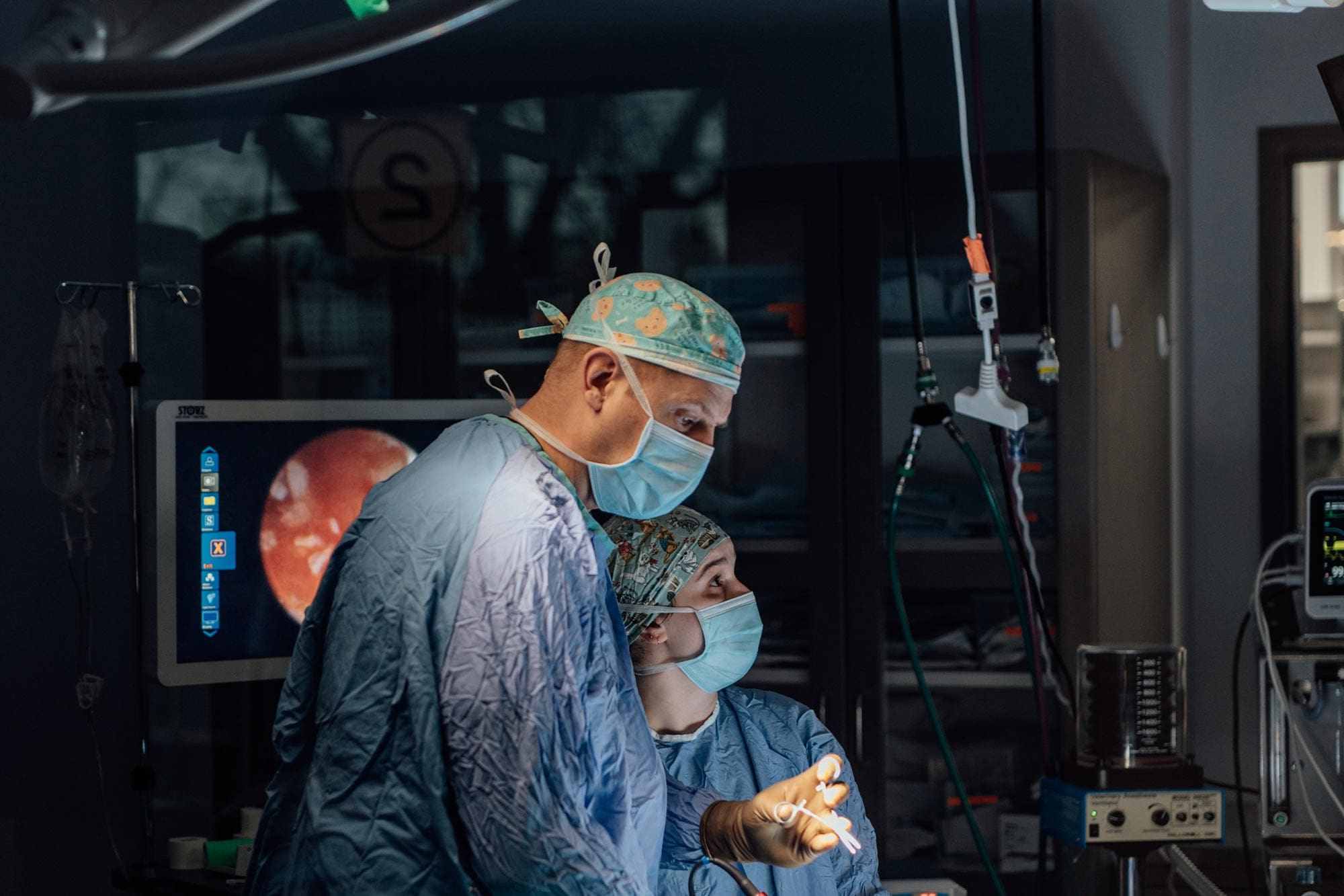
Surgery is the most effective long-term treatment for laryngeal paralysis, with the tie-back procedure being the most common. This involves:
-
Making an incision on the left side of the neck to access the laryngeal cartilages.
-
Identifying the arytenoid cartilage.
-
Using two sutures to permanently hold open one half of the larynx.
-
Closing the incision and confirming the airway is open.
Postoperative Care


-
Prevent your pet from scratching the incision and inspect it daily for swelling, drainage, or suture loss.
-
Clean the area gently with warm water as needed.
-
Schedule suture removal in 10 to 14 days.
-
Limit barking and activity for the first 3 weeks, and use a harness instead of a collar when walking.
-
Hand-feed your pet for the first 1 to 3 weeks until they can eat comfortably without coughing.
-
Avoid swimming post-surgery, as the airway is permanently open.
Potential Complications
Complications from laryngeal tie-back surgery are rare but can include:
-
Long-term coughing or changes in bark or meow
-
Seroma formation of a harmless fluid-filled pocket that typically resolves on its own
-
Aspiration pneumonia that can often be effectively treated when caught early


Prognosis
The outlook for dogs undergoing surgical repair for laryngeal paralysis is generally good. Approximately 85% of dogs experience a significant improvement in breathing and activity levels, leading to a better quality of life. Those with underlying health issues may also have a positive prognosis, depending on their conditions.

