
What is Patellar Luxation in Pets?
The patella, or kneecap, is a small bone located at the junction of the thigh's quadriceps muscles and the tendon that connects to the tibia (shinbone). Its primary role is to facilitate knee function and maintain leg alignment. Patellar luxation occurs when the kneecap dislocates, leading to abnormal cartilage wear, osteoarthritis, and stress on other knee ligaments.
While dogs of any age or breed can experience this condition, it is especially common in small and toy breeds, with about 80% of cases being congenital. Around 50% of dogs with patellar luxation have it in both knees, although one may be more severely affected. The condition typically worsens over time.
Under normal circumstances, the patella glides within a groove at the end of the femur (thigh bone). In cases of patellar luxation, the kneecap rides outside its groove when the knee is bent. This condition can be classified based on whether the patella shifts to the inner (medial) or outer (lateral) side of the knee and whether the luxation is temporary or permanent. Cats can also experience patellar luxation.
Grades of Patellar Luxation
Grade I
The patella is unstable but remains in the groove most of the time. Pets may show lameness during activity but are usually not lame otherwise.
Grade II
The patella shifts out of position but can return to the correct spot on its own. Lameness may become more frequent as the condition progresses.
Grade III
The patella typically stays luxated but can be manually repositioned. Lameness is common, and these patients may suffer from cranial cruciate ligament (CCL) tears.
Grade IV
The patella remains luxated permanently, often causing a crouched walking position. CCL tears are frequently associated with this grade.
Consequences of Untreated Patellar Luxation
If untreated, every instance of the kneecap riding out of its groove can damage cartilage, leading to osteoarthritis and pain. As the trauma continues, the kneecap may increasingly fail to return to its proper position, resulting in chronic lameness and severe osteoarthritis. The rotation caused by patellar luxation can also contribute to CCL tears, worsening lameness over time.
Not all cases require surgical intervention, and your veterinarian will discuss the best treatment options. While physical therapy can strengthen surrounding muscles, it won’t correct poor anatomical alignment.


Surgical Procedures for
Patellar Luxation
Surgical Procedures for Patellar Luxation
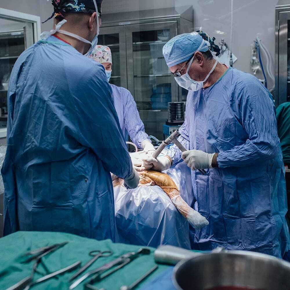
Surgical correction is often necessary for patients experiencing lameness due to patellar luxation. Most cases can be corrected using various techniques, including:
In more severe cases, correction of abnormally shaped femurs and tibias may be required, involving cutting the bone, correcting its shape, and stabilizing it. Generally, higher-grade luxations necessitate more complex procedures and longer recovery times.


Postoperative Prognosis
About 95% of pets achieve good knee function, regain strength and range of motion, and live normal lives. Lower-grade luxations involve simpler procedures and yield better outcomes, while Grade IV luxations may require extensive physical therapy for optimal recovery. Osteoarthritis may progress even after surgery but usually does not cause serious issues later in life.
Postoperative Care
Your pet’s surgeon will create a personalized postoperative care plan, typically involving pain medications for the first 1 to 2 weeks and suture removal 10 to 12 days after surgery.
-
Weeks 1 to 6
Focus on rest, short leashed walks, and light physical therapy, with a follow-up exam at 6 weeks.
-
Weeks 6 to 12
Gradually increase activity, with physical therapy often recommended to help your pet adjust to a straight leg.

