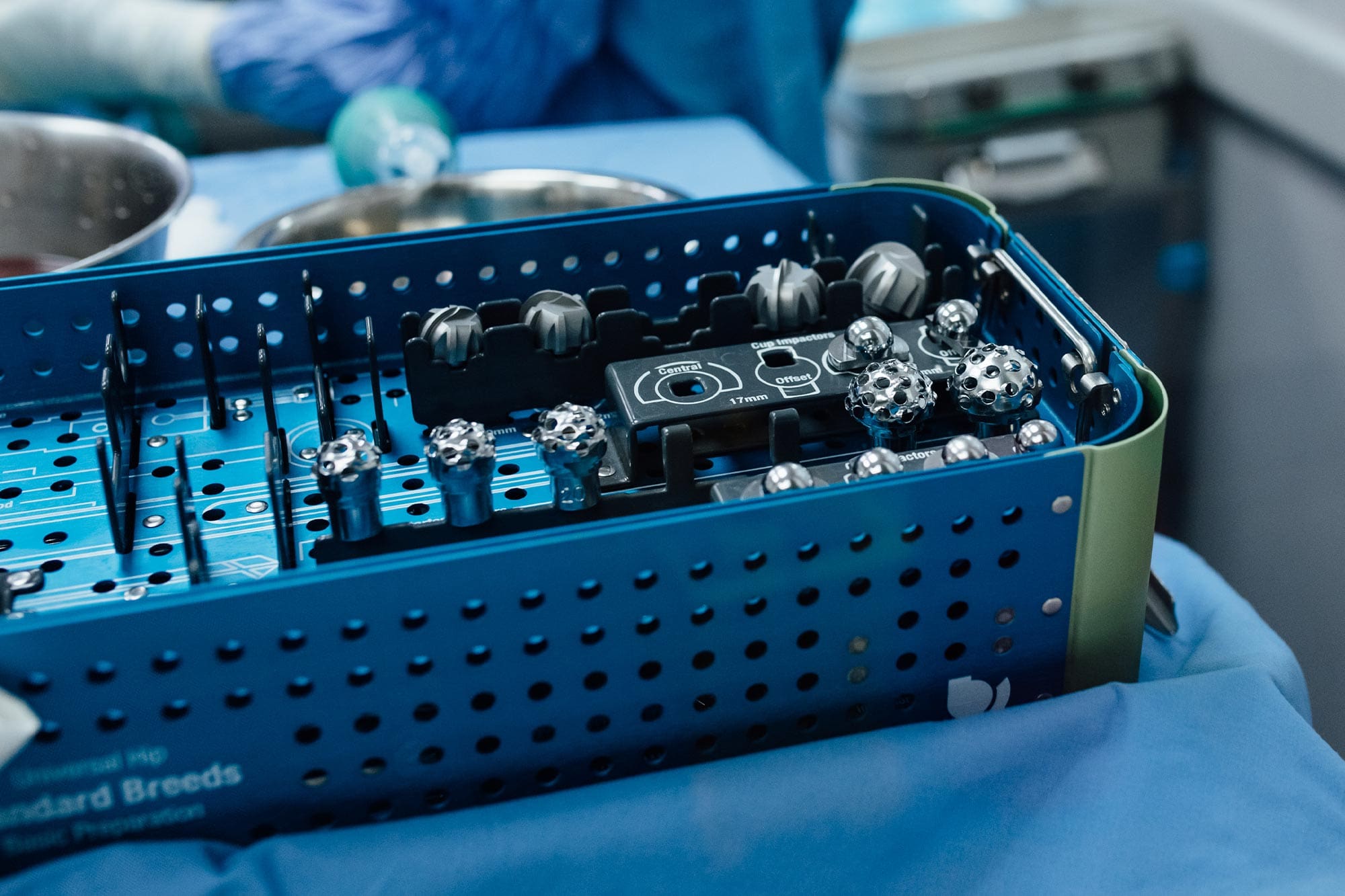
Orthopedic Procedures
Orthopedic surgery addresses injuries, abnormalities, and diseases of the bones, joints, muscles, and ligaments. At Veterinary Specialty Center, our board-certified surgeons combine advanced diagnostics, innovative treatments, and state-of-the-art technology to restore mobility, reduce pain, and improve quality of life for your pet. From minimally invasive therapies to complex surgical reconstructions, we’re here to support you and your pet every step of the way.


Pre-Surgical Workup & Postoperative Recovery
Successful orthopedic care begins with a comprehensive pre-surgical workup and extends through a carefully managed recovery period. Our collaborative team of specialists—including radiologists, surgeons, anesthesiologists, and critical care veterinarians—works together to ensure your pet receives the best care possible.
Pre-surgical diagnostics help us identify the root cause of your pet’s condition and evaluate any underlying health concerns, such as heart or liver disease, that could affect surgery or recovery. These insights allow us to minimize risks and create a treatment plan tailored to your pet’s unique needs.
Following surgery, your pet’s recovery is carefully monitored by our critical care specialists. Rehabilitation therapy and follow-up care through our orthopedic, internal medicine, or primary care services will help your pet regain strength, mobility, and comfort.
Advanced Diagnostics
Diagnosing orthopedic conditions in animals can be challenging—they can’t tell us where it hurts! That’s why we use a combination of physical exams and cutting-edge diagnostic tools to pinpoint the problem. These include:
-
64-Slice Computer-Assisted Tomography (CAT scan)
-
3 Tesla Magnetic Resonance Imaging (MRI)
-
Advanced digital radiography
-
Fluoroscopy
-
3D printing
-
Diagnostic arthroscopy (minimally invasive joint examination)
-
Arthrocentesis (joint fluid analysis)
-
Musculoskeletal ultrasound
-
Video gait analysis
These tools enable us to accurately diagnose the condition and plan the most effective treatment for your pet.
Non-Surgical Treatment Options
In many cases, orthopedic issues can be treated without surgery. Our specialists focus on restoring function and reducing pain through advanced, minimally invasive therapies. These include:
-
Regenerative medicine
-
Platelet-rich plasma (PRP) therapy
-
Stem cell therapy
-
Rehabilitation therapy
-
Integrative therapy
These options may be used alone or alongside other treatments to provide the best outcomes for your pet.
Orthopedic Surgical Options
When surgery is the best option for your pet, our skilled surgeons use advanced techniques and equipment to provide exceptional care. Below are some of the most common conditions we treat:


-
Bone cancer
-
Cranial cruciate ligament injury (CCL)
-
Developmental and juvenile disorders
Including elbow dysplasia, growth deformities, and hip dysplasia
-
Fractures and joint dislocations
-
Limb deformity correction
-
Medial shoulder instability
-
Osteoarthritis
-
Osteochondritis dissecans (OCD)
-
Patellar luxation
-
Shoulder disorders and tendon injuries
-
Traumatic tendon injuries
-
Total hip replacement
Our team also handles rare or complex orthopedic cases. If your pet requires specialized care, don’t hesitate to reach out for more information.