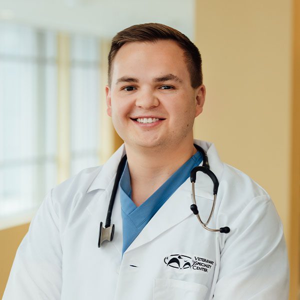
Tim Grote DVM (Resident)
Diagnostic Imaging

DVM
University of Illinois College of Veterinary Medicine
Internship
Veterinary Specialty Center
Dr. Timothy Grote chose a career in veterinary medicine because of his profound passion for animals and a deep fascination for medical science. He finds great joy in caring for their well-being and ensuring they lead healthy, fulfilling lives. His desire to make a positive impact on the lives of animals and their owners is rooted in VSC’s belief that every creature deserves compassionate and expert care. By combining his passion for animals with his keen interest in medical science, he has chosen a path that allows him to make a meaningful difference in the lives of both animals and their human companions.
Get to Know the Expertise, Skill, and Heart Behind VSC
How did you become interested in diagnostic imaging?
I was drawn to the field of veterinary radiology because it plays a crucial role in helping veterinarians make accurate diagnoses and develop effective treatment plans for their patients. I find immense satisfaction in being part of a specialized team that collaborates with veterinarians, using advanced imaging technology to uncover hidden clues and provide critical information that can lead to better outcomes for animals.
What are some of the biggest challenges in your area of expertise?
My interest in veterinary radiology is also fueled by my curiosity to explore and understand the intricacies of animal anatomy, physiology, and pathology through the lens of diagnostic imaging, which is a highly complex field to understand. I appreciate the challenge of interpreting complex images and recognizing subtle abnormalities that may have significant implications for an animal’s health.
Is there a particular case that has inspired you?
I had an obstructive foreign body case in ER that the dog was vomiting and having diarrhea for a week. We diagnosed something that appeared like a toy on ultrasound, and eventually, we tried performing an endoscopy; after significant searching of the stomach and upper small intestine, there was no foreign body. We took him back for an X-ray and ultrasound, but after diagnostics, it still appeared in the stomach. We then took him BACK to endoscopy, and eventually found a corn cob that was lodged behind the scope in a fold of the stomach! Through the radiologist’s meticulous imaging techniques and skills, we saved the pup from going to surgery, and the dog ended up doing great.
What do you like to do outside of work?
I enjoy relaxing with friends, woodworking, golfing, and grilling.
