Diagnostic Equipment
Computed Tomography (CT Scan)
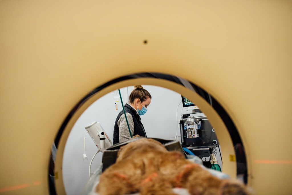 VSC’s 64-slice Computed Tomography (CT) is faster and provides better images than our previous 8-slice CT. It allows us to acquire and reconstruct thin, cross-sectional images (also called slices) of a body part, eliminating the superimposition of structures that occur in conventional radiography. CT is also much more sensitive to differences in tissue density when compared to radiology. It is usually the imaging modality of choice for evaluating bony lesions but can also be used to evaluate soft tissue structures.
The basic technology behind CT involves an x-ray tube that turns around the patient within a gantry. Recent developments in CT have brought a technology called multi-detector scanning. This allows the acquisition of 2, 4, 8, 16 or more slices simultaneously depending on how many rows of detectors the scanner has. It offers the capability of creating thinner or thicker sections (slices) from the same raw data and consequently 3D reconstruction with minimal artifacts.
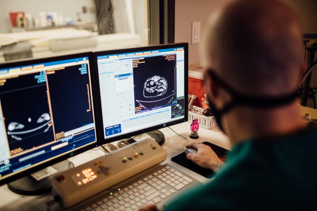
Since acquiring a multi-detector CT scanner, we have been able to provide high-resolution images much faster than previously, which reduces the time patients must spend under general anesthesia.
We commonly perform CT scans of the following:
- Thorax
- Abdomen
- Head
- Bulla
- Nasal cavity
- Neck
- Pelvis
Digital Radiology and Fluoroscopy
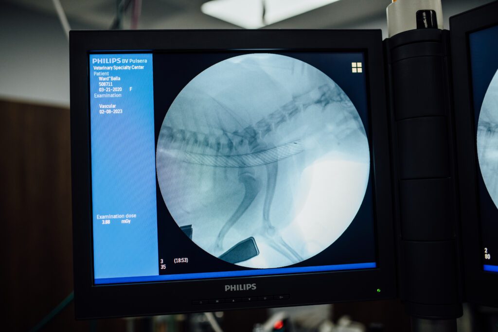 Veterinary Specialty Center has two digital radiology suites for diagnostic radiology and one Fluoroscopy suite for contrast and interventional procedures.
Our entire radiology department is filmless which means that all radiographic studies (XRays) are digital and transferred to our PACS system. Digital radiography has superior image, increases efficiency in daily workflow, decreases exposure of personnel to ionizing radiation, and allows us to integrate all imaging studies for each patient.
Fluoroscopy is a moving x-ray. This moving image is transmitted to a monitor so that the body part and motion can be seen in detail. The C-arm fluoroscopy unit is used to perform dynamic studies, including evaluation for tracheal collapse, swallowing dysfunction, and intravenous pyelography (IVP) for ectopic ureters. It is also used for interventional cardiology procedures. The unit can be rotated around the patient to obtain different views without changing the position of the animal.
Interventional Radiology
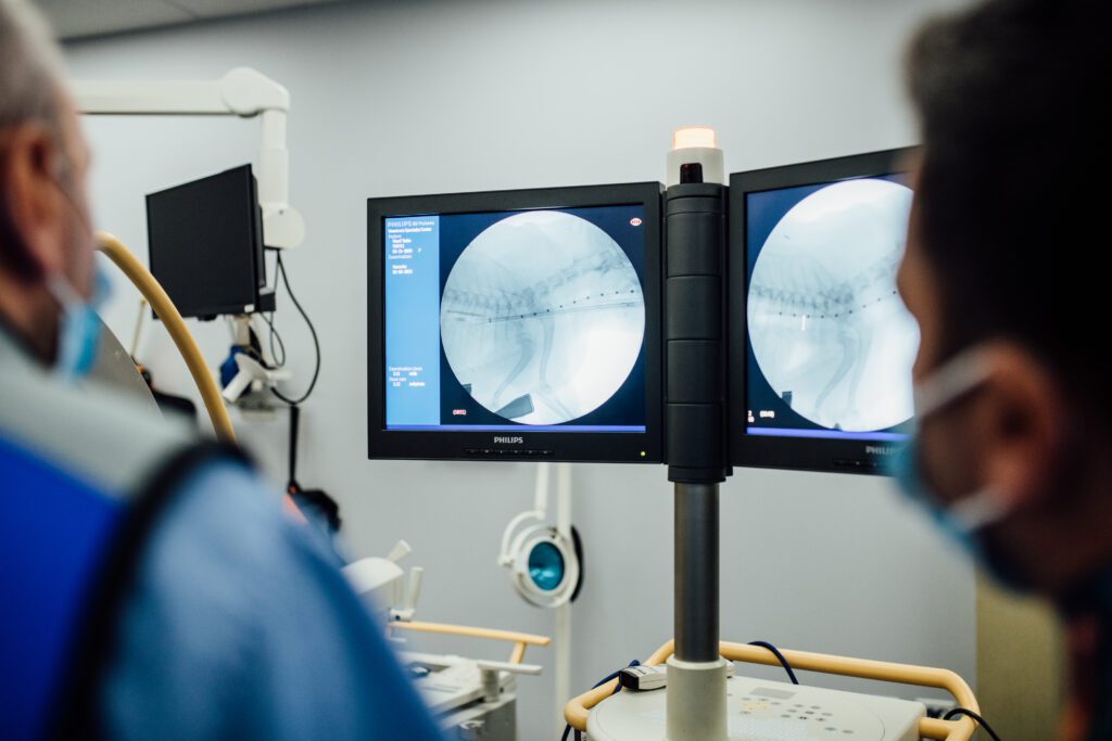 Interventional Radiology involves the use of fluoroscopy, ultrasound, CT, MRI and endoscopy, (or various combinations of these) to gain access to different structures in order to deliver materials for therapeutic purposes. These procedures are minimally invasive and less painful. In addition, it offers treatment options for patients with various conditions that cannot be managed or treated with surgery, or that are associated with excessive morbidity, high cost or poor outcome. It requires multiple guide wires with various properties, catheters specifically adapted for individual procedures, stents composed of different materials and configurations, embolic coils, embolic particles, drainage devices, glue, oils, chemotherapeutic agents, occlusion devices, or balloons.
We have the equipment and specialist expertise at VSC to perform the following procedures:
- Tracheoplasty (tracheal stenting)
- Intrahepatic portosystemic shunt embolization
- Chemoembolization of hepatic tumors
- Urethral stenting
- Embolization of arterio-venous malformations
- Vascular nasal embolization of intractable epistaxis
- Vascular foreign body retrieval
- Percutaneous nephrostomy tube placement
- Antegrade placement of urinary catheter
- Vascular stenting and angioplasty
- Thrombolytic vascular procedures
Magnetic Resonance Imaging (MRI)
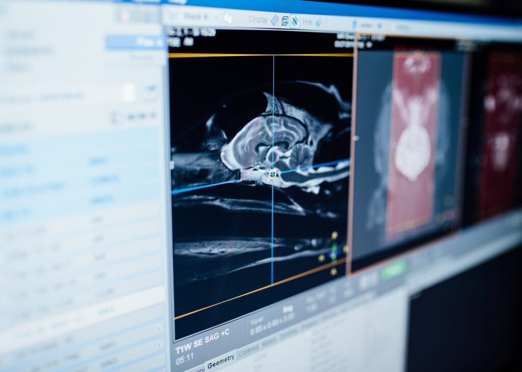
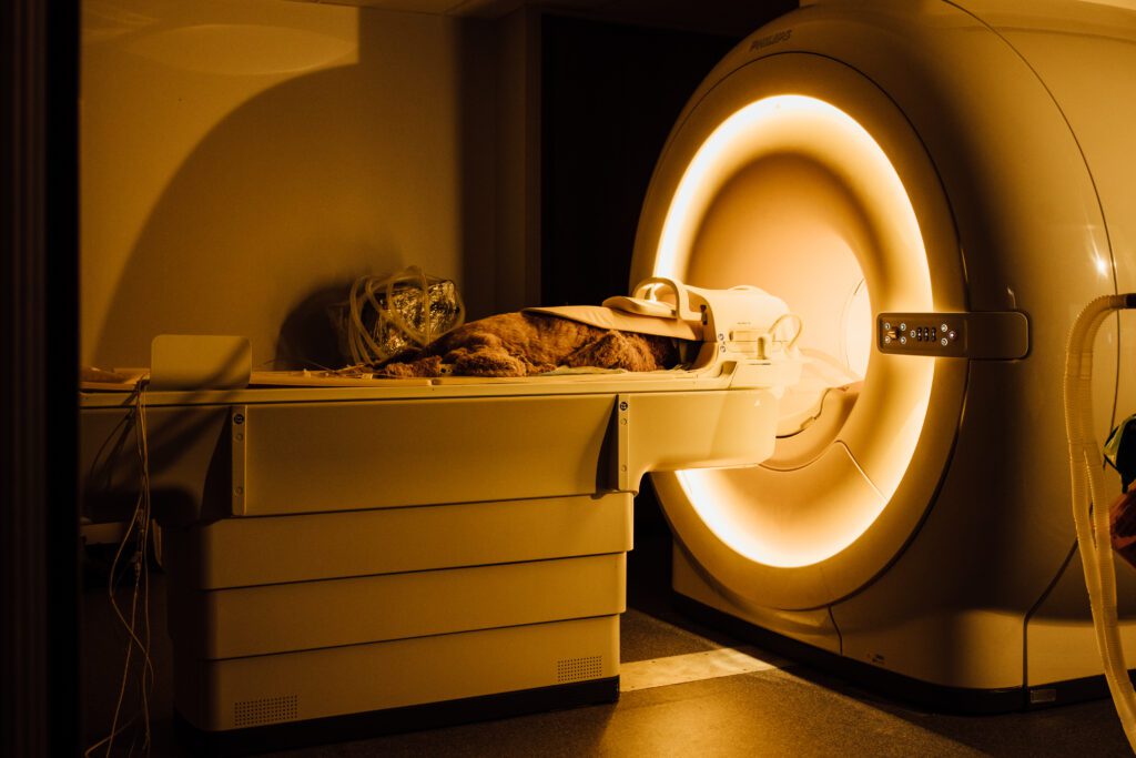
In October of 2018, VSC upgraded our Magnetic Resonance Imaging (MRI) from a 1 Tesla to a 3 Tesla. Our new MRI is three times stronger than our previous MRI unit and gives us top-of-the-line image quality as well as faster scan times, reducing the anesthesia time for our patients. Because the detail is so much better than in the past, we can examine smaller areas of the body, smaller animals and musculoskeletal structures such as the shoulder and get great resolution. VSC is one of only a few veterinary hospitals in a non-university setting to have a 3 Tesla MRI. It is presently the only 3 Tesla MRI in a veterinary setting in the Chicago area.
Magnetic Resonance Imaging (MRI) uses a magnetic field and radio waves to create a detailed picture of the organs and tissues within your pet’s body. An MRI produces high-resolution images of the inside of the body that helps diagnose a variety of problems. Unlike radiographs and CT Scans, an MRI does not use radiation.
An MRI is used to provide clear images of parts of the body that can’t be seen as well on a CT scan, radiographs or ultrasound. The ability to highlight contrast in soft tissue makes an MRI useful to visualize the brain, spinal cord and other soft tissues. A pet receiving an MRI is anesthetized and monitored closely in the MRI machine.
The most common applications for MRI are:
- Neurologic disease (brain and spinal cord)
- Evaluation of tumor margins
- Occult lameness
Nuclear Scintigraphy
VSC is equipped with a digital gamma camera for nuclear scintigraphy, which allows the functional evaluation of different organs or systems. It uses small, tracer amounts of radioactive molecules to diagnose diseases involving bone, soft tissue and vessels and can often diagnose diseases not visible with other imaging methods.
The most common applications are:
Thyroid Scans: These are used primarily to confirm the presence of hyper functional tissue in cats that are suspected to be hyperthyroid and to help evaluate the origin of neck masses in dogs.
Bone scans: Bone scans are used primarily in cases of occult lameness or to localize metastatic bone lesions. Bone scans allow us to evaluate the patient’s entire skeleton in a cost and time-efficient manner.
PACS Teleradiology
 All of our imaging modalities are digitized and linked to a Picture Archiving and Communication System (PACS) where all images are digitally available. This system enables members of our teams to be able to retrieve imaging studies in a matter of seconds to easily compare them to each other.
Veterinary Specialty Center has 18 PACS viewing stations, which allows specialists throughout the hospital to have quick access to all of our patients’ images. It also allows us to make CDs or email images to you and your regular veterinarian. This facilitates discussion of cases, as our specialists and your regular veterinarian can consult the images together from different locations.
Ultrasonography or Ultrasounds
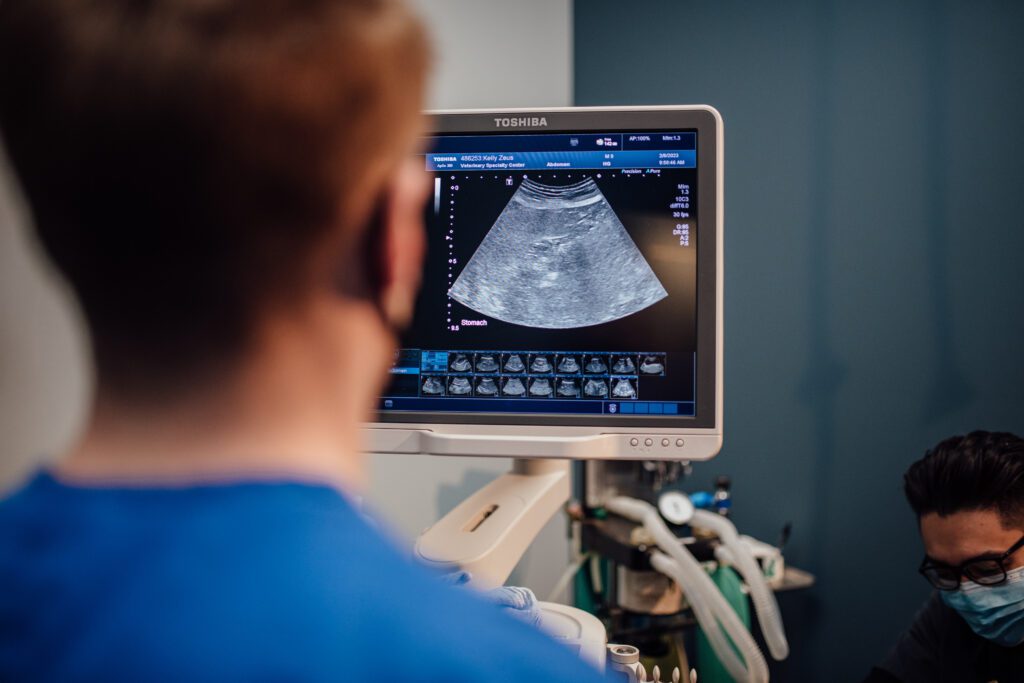 Ultrasonography or an ultrasound, uses high-frequency sound waves to produce pictures inside your pet’s body. Because the images are captured in real-time, they can show the structure and movement of the body’s internal organs as well as the flow of blood through blood vessels. It is used primarily to evaluate soft tissue structures. The most common ultrasound examination that we perform is an abdominal ultrasound, where we evaluate all of the organs within the abdomen (from the liver to the urinary bladder).
Other examinations that we commonly perform include:
Thoracic ultrasonography
The ultrasound beam cannot pass through air and so cannot be used to evaluate normal lungs. However, ultrasound can be used to evaluate for lung tumors or to evaluate for lesions within the pleural space or the mediastinum. Ultrasound can also be used to evaluate the heart.
Cervical ultrasonography
We routinely evaluate the thyroid and parathyroid glands, the salivary glands, and the lymph nodes.
Musculoskeletal ultrasonography
Although any soft tissue structure can be evaluated (including tendons, ligaments, muscle, and masses), the most common application is the shoulder for bicipital tendonitis.
We are also able to perform ultrasound-guided aspirates and biopsies of lesions in order to obtain a definitive diagnosis.
